")
Back to Journals » International Journal of Nanomedicine » Volume 9 » Issue 1
Biosynthesis and characterization of silver nanoparticles using panchakavya, an Indian traditional farming formulating agent
Authors Govarthanan M , Selvankumar T, Manoharan K, Rathika R, Shanthi R, Lee K, Cho M, Kamala-Kannan S, Oh B, Chinnapan S, Arumugam S, Loganathan P
Received 9 December 2013
Accepted for publication 22 January 2014
Published 31 March 2014 Volume 2014:9(1) Pages 1593—1599
DOI https://doi.org/10.2147/IJN.S58932
Checked for plagiarism Yes
Review by Single anonymous peer review
Peer reviewer comments 4
Muthusamy Govarthanan,1,2 Thangasamy Selvankumar,2 Koildhasan Manoharan,3 Rajiniganth Rathika,2 Kuppusamy Shanthi,4 Kui-Jae Lee,1 Min Cho,1 Seralathan Kamala-Kannan,1 Byung-Taek Oh1
1Division of Biotechnology, Advanced Institute of Environment and Bioscience, College of Environmental and Bioresource Sciences, Chonbuk National University, Iksan, South Korea; 2PG and Research Department of Biotechnology, Mahendra Arts and Science College, Kalippatti, Namakkal, Tamil Nadu, India; 3Department of Botany, Raja Duraisingam Government Arts College, Sivagangai, Tamil Nadu, India; 4Department of Environmental Science, PSG College of Arts and Science, Coimbatore, Tamil Nadu, India
Abstract: Synthesis of silver nanoparticles (AgNPs) with biological properties is of vast significance in the development of scientifically valuable products. In the present study, we describe simple, unprecedented, nontoxic, eco-friendly, green synthesis of AgNPs using an Indian traditional farming formulating agent, panchakavya. Silver nitrate (1 mM) solution was mixed with panchakavya filtrate for the synthesis of AgNPs. The nanometallic dispersion was characterized by surface plasmon absorbance measuring 430 nm. Transmission electron microscopy showed the morphology and size of the AgNPs. Scanning electron microscopy–energy-dispersive spectroscopy and X-ray diffraction analysis confirmed the presence of AgNPs. Fourier transform infrared spectroscopy analysis revealed that proteins in the panchakavya were involved in the reduction and capping of AgNPs. In addition, we studied the antibacterial activity of synthesized AgNPs. The synthesized AgNPs (1–4 mM) extensively reduced the growth rate of antibiotic resistant bacteria such as Aeromonas sp., Acinetobacter sp., and Citrobacter sp., according to the increasing concentration of AgNPs.
Keywords: green synthesis, panchakavya, antibacterial, environment, nanoparticles
Introduction
Synthesis of nanoparticles (NPs) has received considerable attention in recent years due to the wide range of NP applications in different scientific fields, such as medicine, biotechnology, chemistry, physics, catalysis, electronics, and materials science.1,2 The metallic NPs exhibit numerous physical and chemical properties that are not observed either in the individual molecules or in the bulk materials.3 Among the metallic NPs, silver NPs (AgNPs) in particular are known for their versatile biological applications in the fields of medicine and biotechnology.4,5 AgNPs can be synthesized by physical and chemical methods.6–11 The chemical synthesis of AgNPs may lead to the presence of some toxic chemicals adsorbed on the surface that may have adverse effects in its application and also the chemical used in the synthesis may pollute the environment.5
Biological materials have the potential to reduce the metal ions into metal NPs. Biosynthesis of AgNps gained considerable attention in the past decade. Moreover, it has been proposed as a less toxic, cost-effective, environmentally friendly alternative to chemical and physical methods. Accordingly, AgNPs have been synthesized using microorganisms,12–15 plant extracts,16 and milk.17 The proteins present in the biological materials are involved in the reduction and stabilization of NPs.18
Livestock plays a vital role in the income of Indian farmers and livestock products have a significant place in the livelihoods of farmers. For sustainable agricultural management, Indian farmers have developed several traditional formulations based on their knowledge accumulated through generations of experience. Panchakavya is one such excellent formulation, consisting of five products from cows: milk, curd, ghee, urine, and dung.19 Principally, panchakavya is a mixture of microorganisms, including Azospirillum sp., Azatobacter sp., Pseudomonas sp., and many other beneficial organisms. Moreover, it also contains proteins, lipids, carbohydrates, micronutrients, and antioxidants.19 However, there has been no report to date on the synthesis of AgNPs using panchakavya.
The antibiotic-resistant Citrobacter sp. and Aeromonas sp. are Enterobacteriaceae-family clinical pathogens that cause a wide range of infections, including urinary tract infections, neonatal sepsis, meningitis, bloodstream infections, and pneumonia.20–22 Acinetobacter sp., which belongs to the Gram-negative Gammaproteobacteria class, causes a wide spectrum of infections that include pneumonia, bacteremia, meningitis, urinary tract infection, and wound infection.22,23 The high mortality rate associated with these microbial infections may be due in part to ineffective empirical antibiotic therapy. These microorganisms are commonly resistant to extended-spectrum antibiotics.23 However, the populations of antibiotic-resistant microorganisms are continuously increasing, and their control is a major challenge for scientists and researchers.24 The wastes discharged from pharmaceutical industries, hospitals, and research laboratories contain different groups of antibiotics and other pollutants. The presence of antibiotics in the ecosystem induces the development of an antibiotic resistance mechanism in microbial communities. Hence, the synthesis of AgNPs and their antimicrobial properties are emerging as areas of great interest among researchers. AgNPs have been widely studied for their antibacterial,25 antifungal,17 and antiviral26,27 properties due to their large surface area. Accordingly, the objectives of the present study were 1) to synthesize AgNPs using panchakavya extract; 2) characterization of synthesized AgNPs; and 3) evaluation of the antibacterial activity of biologically synthesized AgNPs.
Materials and methods
Panchakavya
Panchakavya was procured from Sakthi Vermicomposting farm, Madurai, Tamil Nadu, India. The principal constituents of panchakavya are presented in Table 1. The mixture of panchakavya was filtered through Whatman No 1 filter paper followed by 0.2 μm membrane filter, and the filtrate was used for the synthesis of AgNPs.
 | Table 1 Principal constituents of panchakavya |
Synthesis of AgNPs
AgNO3 was purchased from Sigma-Aldrich (St Louis, MO, USA), and the synthesis procedure was carried out according to the methods of Lee et al.17 Briefly, 4 mL of panchakavya was mixed with 96 mL of 1 mM AgNO3 solution, and the resulting milky-white mixture was incubated for 8 hours in a rotary shaker (180 rpm) at room temperature. Reduction of Ag+ ions to Ag was monitored by change in color of the reaction mixture from milky white to dark brown.
Characterization techniques
The synthesis of AgNPs was characterized preliminarily by scanning the absorption maxima of the mixture at wavelengths between 300–700 nm on a UV-1800 ultraviolet-visible (UV-Vis) spectrophotometer (Shimadzu, Kyoto, Japan). The characterization of the synthesized AgNPs was conducted with a Rigaku X-ray diffractometer (Tokyo, Japan), operated at 2θ from 30° to 80° at 0.041°/minute with a time constant of 2 seconds. AgNP synthesis and the elemental composition were further confirmed by scanning electron microscopy–energy dispersive spectroscopy ([SEM–EDS] JEOL-64000; JEOL, Tokyo, Japan). The surface morphology, crystalline nature, and size of the synthesized AgNPs were examined using high-resolution transmission electron microscopy ([TEM] JEM-2010; JEOL) at an accelerating voltage of 200 kV. The spectra of the AgNPs were obtained by Fourier transform infrared spectroscopy ([FTIR] PerkinElmer, Waltham, MA, USA) in the diffuse reflectance mode at a resolution of 4 −1 with KBr pellets.
Antibacterial activity
The antibiotic-resistant bacterial strains Citrobacter sp., Acinetobacter sp., and Aeromonas sp. were allowed to grow in 100 mL of Luria-Bertani broth supplemented with synthesized AgNPs ranging from 1–4 mM. The growth of the bacterial strains was indexed by measuring the optical density (at λ=600 nm) at regular intervals (0–48 hours) using the Shimadzu UV-1800 spectrophotometer. The growth curve was plotted between optical density and time.
Results and discussion
The reduction of AgNO3 using biological materials is the most widely practiced method for the green synthesis of AgNPs in colloidal form. After 8 hours’ incubation at room temperature, the milky-white reaction mixture turned into a yellowish brown color. The appearance of yellowish brown color is the indication of AgNO3 reduction. The biomolecules present in panchakavya may be involved in the reduction of AgNO3 to AgNPs. A recent report of AgNPs synthesis has established the significant role of proteins in reduction of AgNO3,13 and Litvin et al28 reported the formation of AgNPs using humic substance. In the present study, panchakavya was mixed in an aqueous solution of silver ion complex and the reduction of pure Ag+ ions to Ag0 was monitored by measuring the UV-Vis spectrum of the reaction media. Figure 1 shows that the UV-Vis spectra of the silver surface plasmon resonance band occurs near 430 nm. The results are consistent with a previous report.18 The samples were subjected to TEM, X-ray diffraction (XRD) and SEM–EDS to confirm the presence of Ag. TEM images of the biosynthesized AgNPs showed that the particles were spherical in shape with an average size of 20 to 100 nm (Figure 2). To further validate the AgNPs, the samples were analyzed in SEM–EDS and the results are shown in Figure 3. The results show strong silver signals at 3 keV. The EDS quantitative analysis showed the presence of silver (100%) without any contaminants. FTIR is an important tool for the identification of functional groups and interactions between molecules.29 The bands observed at 3,416 and 2,920 cm−1 were assigned to the C-H stretching vibrations of the primary and secondary amines, respectively. The sharp absorption peaks at 1,629 and 1,046 cm−1 (Figure 4) could be of N–H stretching in primary and secondary amines and amides.18 The crystalline nature of the synthesized AgNPs was investigated by XRD and the corresponding XRD diffractogram is shown in Figure 5. The XRD peaks at 46.1°, 54.55°, 67.74°, and 76.84° correspond to 111, 200, 142, 220, and 311 planes for silver (JCPDS card number 04-0783).5,17
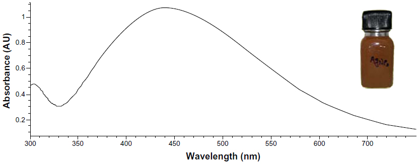 | Figure 1 Ultraviolet-visible absorption spectrum of the silver nanoparticles prepared from 1 mM silver nitrate solution. |
 | Figure 2 Transmission electron microscopy image of silver nanoparticles. |
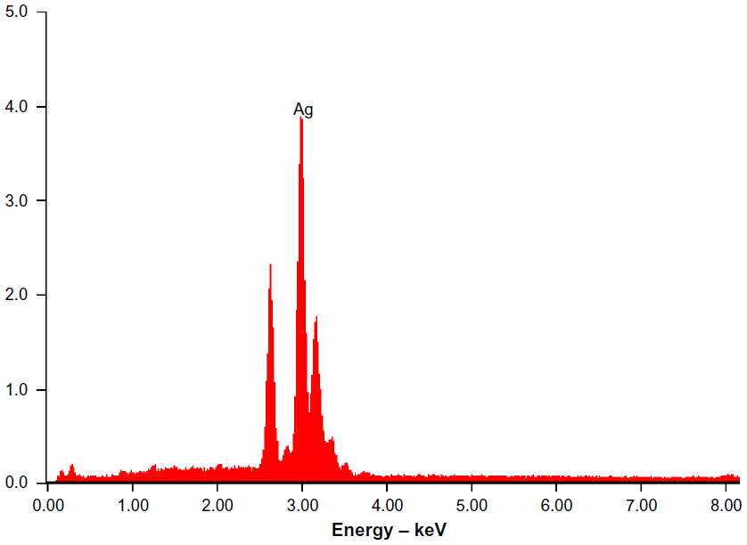 | Figure 3 Scanning electron microscopy–energy dispersive spectrum of silver (Ag) nanoparticles. |
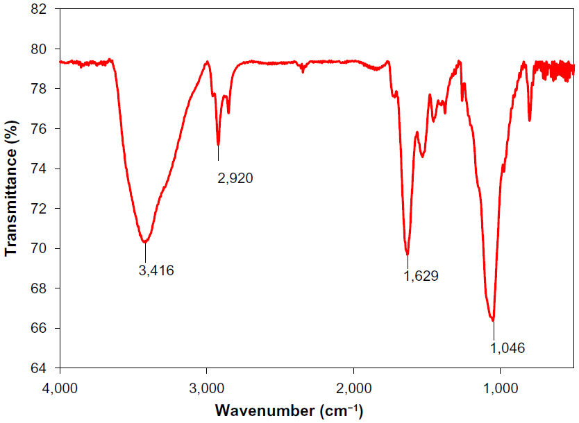 | Figure 4 Fourier transform infrared spectra of purified silver nanoparticles synthesized from panchakavya. |
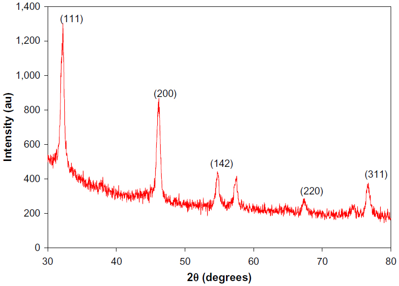 | Figure 5 X-ray diffraction pattern of silver nanoparticles synthesized using panchakavya. |
Biosynthesized AgNPs were studied for antibacterial activity against antibiotic-resistant bacteria, namely, Citrobacter sp., Acinetobacter sp., and Aeromonas sp. The bacterial growth in different concentrations (1–4 mM) of AgNPs was determined and plotted as a function of time for 0–48 hours at regular intervals (Figure 6). The results indicate that 4 mM of AgNPs effectively inhibited the bacterial growth. In general, the growth rate of the bacterial population was decreased according to the increasing concentration of AgNPs. The results are consistent with studies reporting the antibacterial activity of AgNPs.30–32
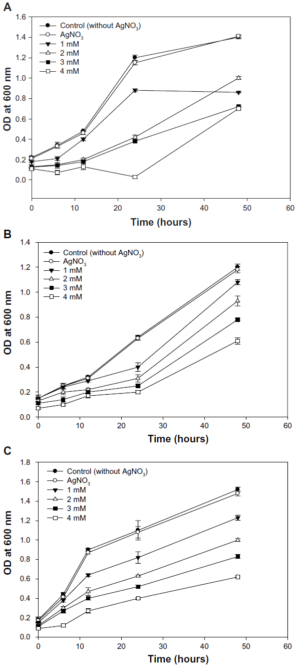 | Figure 6 Effect of silver nanoparticles on growth of antibiotic-resistant bacteria. |
Conclusion
We used nontoxic, eco-friendly panchakavya for the consistent and rapid synthesis of AgNPs. The synthesized particles were circular in shape and the elemental composition confirmed the presence of silver without any contamination. The prepared AgNPs had significant antibacterial activity against antibiotic-resistant bacteria. Application of AgNPs based on this conclusion may lead to diverse applications in the fields of science and technology.
Acknowledgments
This research was supported by the Korean National Research Foundation (Korean Ministry of Education, Science and Technology, Award NRF-2011-35B-D00020). This research work was partially supported by the National Research Foundation of Korea (NRF) grant funded by the government (MEST; No 2011-0020202). This work was also partially supported by Korea Ministry of Environment as “The GAIA Project (No 2012000550021).” The authors are grateful to Mr S Sivasami, Sakthi Vermicomposting farm, Sanampatti, Madurai, Tamil Nadu, India for supplying the panchakavya.
Disclosure
The authors report no conflicts of interest in this work.
References
Raveendran P, Fu J, Wallen SL. Completely “green” synthesis and stabilization of metal nanoparticles. J Am Chem Soc. 2003;125(46):13940–13941. | |
Prow T, Smith JN, Grebe R, et al. Construction, gene delivery, and expression of DNA tethered nanoparticles. Mol Vis. 2006;12:606–615. | |
Daniel MC, Astruc D. Gold nanoparticles: assembly, supramolecular chemistry, quantum-size-related properties, and applications toward biology, catalysis, and nanotechnology. Chem Rev. 2004;104:293–346. | |
Becker RO. Silver ions in the treatment of local infections. Met Based Drugs. 1999;6:311–314. | |
Suman TY, Radhika Rajasree SR, Kanchana A, Elizabeth SB. Biosynthesis, characterization and cytotoxic effect of plant mediated silver nanoparticles using Morinda citrifolia root extract. Colloids Surf B Biointerfaces. 2013;106:74–78. | |
Huang H, Yang Y. Preparation of silver nanoparticles in inorganic clay suspensions. Compos Sci Technol. 2008;68:2948–2953. | |
Nadagouda MN, Speth TF, Varma RS. Microwave-assisted green synthesis of silver nanostructures. Acc Chem Res. 2011;44(7):469–478. | |
Ahmad T, Wani IA, Manzoor N, Ahmed J, Asiri AM. Biosynthesis, structural characterization and antimicrobial activity of gold and silver nanoparticles. Colloids Surf B Biointerfaces. 2013;107:227–234. | |
Sastry M, Ahmad A, Khan MI, Kumar R. Biosynthesis of metal nanoparticles using fungi and actinomycete. Curr Sci. 2003;85:162–170. | |
Wani IA, Gangulya A, Ahmed J, Ahmad T. Silver nanoparticles: ultrasonic wave assisted synthesis, optical characterization and surface area studies. Mater Lett. 2011;65:520–522. | |
Wani IA, Khatoon S, Ganguly A, Ahmed J, Ganguli AK, Ahmad T. Silver nanoparticles: large scale solvothermal synthesis and optical properties. Mater Res Bull. 2010;45:1033–1038. | |
Ahmad A, Senapati S, Khan MI, Kumar R, Sastry M. Extracellular biosynthesis of monodisperse gold nanoparticles by a novel extremophilic actinomycete, Thermomonospora sp. Langmuir. 2003;19:3550–3553. | |
Shahverdi AR, Minaeian S, Shahverdi HR, Jamalifar H, Nohi A. Rapid synthesis of silver nanoparticles using culture supernatants of Enterobacteria: a novel biological approach. Process Biochem. 2007;42:919–923. | |
Jha AK, Prasad K, Prasad K. A green low-cost biosynthesis of Sb2O3 nanoparticles. Biochem Eng J. 2009;43:303–306. | |
Bharde AA, Parikh RY, Baidakova M, et al. Bacteria-mediated precursor-dependent biosynthesis of superparamagnetic iron oxide and iron sulfide nanoparticles. Langmuir. 2008;24:5787–5794. | |
Kumar V, Yadav SK. Plant-mediated synthesis of silver and gold nanoparticles and their applications. J Chem Technol Biotechnol. 2009;84:151–157. | |
Lee KJ, Park SH, Govarthanan M, et al. Synthesis of silver nanoparticles using cow milk and their antifungal activity against phytopathogens. Mater Lett. 2013;105:128–131. | |
Velmurugan P, Shim J, Kamala-Kannan S, et al. Crystallization of silver through reduction process using Elaeis guineensis biosolid extract. Biotechnol Prog. 2011;27:273–279. | |
Ganesh Kumar K, Kumaravelu N, Sivakumar T, Gajendran K. Study on panchakavya – an indigenous formulation and its effect on the growth promotion of crossbred pigs. Indian J Anim Res. 2006;40:158–160. | |
Kline MW. Citrobacter meningitis and brain abscess in infancy: epidemiology, pathogenesis, and treatment. J. Pediatr. 1988;113:430–434. | |
Mateos Rodríguez F, Pérez Moro E, Atienza Morales MP, Beato Pérez JL. [Community-acquired bacteremic pneumonia due to Citrobacter diversus]. An Med Interna. 2000;17:165–166. Spanish. | |
Zúñiga J, González P, Henríquez A, Fernández A. [Bacteremic pneumonia caused by Citrobacter diversus: report of a case]. Rev Med Chil. 1991;119:303–307. Spanish. | |
Pepperell C, Kus JV, Gardam MA, Humar A, Burrows LL. Low-virulence Citrobacter species encode resistance to multiple antimicrobials. Antimicrob Agents Chemother. 2002;46:3555–3560. | |
Rai MK, Deshmukh SD, Ingle AP, Gade AK. Silver nanoparticles: the powerful nanoweapon against multidrug-resistant bacteria. J Appl Microbiol. 2012;112(5):841–852. | |
Kim JS, Kuk E, Yu KN, et al. Antimicrobial effects of silver nanoparticles. Nanomedicine. 2007;3:95–101. | |
Sun RW, Chen R, Chung NP, Ho CM, Lin CL, Che CM. Silver nanoparticles fabricated in Hepes buffer exhibit cytoprotective activities toward HIV-1 infected cells. Chem Commun (Camb). 2005:5059–5061. | |
Lu L, Sun RW, Chen R, et al. Silver nanoparticles inhibit hepatitis B virus replication. Antivir Ther. 2008;13:253–262. | |
Litvin VA, Galagan RL, Minaev BF. Kinetic and mechanism formation of silver nanoparticles coated by synthetic humic substances. Colloids Surf A Physicochem Eng Asp. 2012;414:234–243. | |
Kong J, Yu S. Fourier transform infrared spectroscopic analysis of protein secondary structures. Acta Biochim Biophys Sin (Shanghai). 2007;39:549–559. | |
Kasthuri J, Veerapandian S, Rajendiran N. Biological synthesis of silver and gold nanoparticles using apiin as reducing agent. Colloids Surf B Biointerfaces. 2009;68:55–60. | |
Shrivastava S, Bera T, Roy A, Singh G, Ramachandrarao P, Dash D. Characterization of enhanced antibacterial effects of novel silver nanoparticles. Nanotechnology. 2007;18:225103–225112. | |
Litvin VA, Minaev BF. Spectroscopy study of silver nanoparticles fabrication using synthetic humic substances and their antimicrobial activity. Spectrochim Acta A Mol Biomol Spectrosc. 2013;108:115–122. |
 © 2014 The Author(s). This work is published and licensed by Dove Medical Press Limited. The full terms of this license are available at https://www.dovepress.com/terms.php and incorporate the Creative Commons Attribution - Non Commercial (unported, v3.0) License.
By accessing the work you hereby accept the Terms. Non-commercial uses of the work are permitted without any further permission from Dove Medical Press Limited, provided the work is properly attributed. For permission for commercial use of this work, please see paragraphs 4.2 and 5 of our Terms.
© 2014 The Author(s). This work is published and licensed by Dove Medical Press Limited. The full terms of this license are available at https://www.dovepress.com/terms.php and incorporate the Creative Commons Attribution - Non Commercial (unported, v3.0) License.
By accessing the work you hereby accept the Terms. Non-commercial uses of the work are permitted without any further permission from Dove Medical Press Limited, provided the work is properly attributed. For permission for commercial use of this work, please see paragraphs 4.2 and 5 of our Terms.