")
Back to Journals » Clinical, Cosmetic and Investigational Dentistry » Volume 6
A comparative study of retention of complete denture base with different types of posterior palatal seals – an in vivo study
Authors Chandu GS, Hema B, Mahajan H, Azad A, Sharma I, Azad A
Received 29 August 2014
Accepted for publication 15 October 2014
Published 26 November 2014 Volume 2014:6 Pages 95—100
DOI https://doi.org/10.2147/CCIDE.S73446
Checked for plagiarism Yes
Review by Single anonymous peer review
Peer reviewer comments 3
Editor who approved publication: Professor Christopher E. Okunseri
GS Chandu,1 BS Hema,2 Harsh Mahajan,1 Antriksh Azad,2 Ipsita Sharma,3 Anurag Azad4
1Department of Prosthodontics, Rishiraj College of Dental Sciences and Research Centre, Bhopal, Madhya Pradesh, India; 2Department of Conservative Dentistry, Rishiraj College of Dental Sciences and Research Centre, Bhopal, Madhya Pradesh, India; 3Department of Oral Pathology, RKDF Dental college and Research centre, Bhopal, Madhya Pradesh, India; 4Department of Oral and Maxillofacial Surgery, Bhabha College of Dental Sciences and Research Centre, Bhopal, Madhya Pradesh, India
Aim: To evaluate retention of complete denture base with different types of posterior palatal seals.
Material and methods: Ten male patients between the age group of 50 years to 60 years were selected for the study. After the primary and secondary impressions were taken, five casts were made including a cast without posterior palatal seal, a cast with single bead posterior palatal seal, a cast with double bead posterior palatal seal, a cast with butterfly shaped posterior palatal seal, and a cast with posterior palatal seal with low fusing compound by functional method.
Results: It was observed that retention increased up to 108% in the posterior palatal seal with low fusing compound with functional method and the posterior palatal seal that was obtained by using functional method provided greater retention than a denture base without posterior palatal seal.
Conclusion: It was concluded that the incorporation of a posterior palatal seal is important for obtaining optimum retention of the maxillary complete denture.
Keywords: posterior palatal seal, denture base, complete denture, functional method
Introduction
The rationale for the posterior palatal seal lies in completing peripheral seal along the posterior denture border. It provides an effective barrier against the ingress of air or fluid during the functional movement of the soft palate or slight movement of denture during function.1
The available literature states that addition of posterior palatal seal markedly increases the retention of the denture.
To test the retention of maxillary complete denture base both caudally directed intra-oral and cranially directed extra-oral forces have been applied. The following study was carried out for comparative evaluation of retention of complete maxillary denture bases with different posterior palatal seals.
Material and methods
The study was carried out in the Department of Prosthodontics Crown and Bridge of PMNM Dental College and Hospital, Bagalkot, ethical committee reference number 2847. Ten male patients with an average age of 60 years were selected for the study. The selection was based on the clinical examination and evaluation of the diagnostic cast.
Subjects who had been edentulous for varying periods of 6 months to 2 years were selected. The subjects were without any systemic diseases and with good neuromuscular control for better cooperation. The ridges were well-healed, round, smooth, and oral mucosa covering the ridges was firmly attached with average resiliency and thickness. Patients with ridges without bilateral or anterior undercuts were selected. All the subjects had normal salivary flow. The nature of study to be carried out was explained to all subjects.
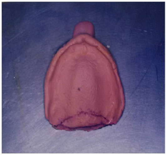
The primary impression was made with an impression compound (Y-dent, Delhi, India) and was border-molded to get the acceptable extension and border thickness (Figure 1). The uniform thickness of a 2 mm spacer was adapted over the cast and keeping it short by 2 mm all over the periphery and a tray was prepared with self-activated acrylic resin (Figure 2). A secondary impression was made with free-flowing zinc oxide eugenol paste (DPI impression paste, DPI, Mumbai, India) with light pressure and border-molding (Figure 3).
 | Figure 1 Primary impression. |
 | Figure 2 Primary cast with special tray. |
 | Figure 3 Secondary impression with zinc oxide eugenol paste. |
The posterior palatal seal area was marked by first locating the pterygomaxillary (hamular) notch with the T burnisher or Moons probe and then marking the line 1.5 mm distal to hamular notches. The posterior palatal seal area marking over the cast was slightly scraped with a sharp instrument to represent the posterior palatal seal area of subsequent casts. The master cast was marked “M”. Dental stone (Dutt stone, 30:100) was poured into the molds over a vibrator to avoid the entrapment of air bubbles. The casts were recovered after 1 hour and marked “A”, “B”, and “C” respectively (Figure 4).
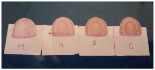 | Figure 4 Duplicate casts. |
On cast “A” a V-shaped groove 1 to 1.5 mm deep and 1.5 mm at its base was carved 2 mm anterior to vibrating line-junction of hard and soft palate. On cast “B” a groove of 1 to 1.5 mm deep and 1.5 mm wide was carved 1 mm anterior to the vibrating line, passing through the hamular notch. An additional groove 1 mm deep and 1 mm wide at its base was carved just distal to anterior limit of posterior palatal seal area. On cast “C” a butterfly shaped design was carved as suggested by Hardy and Kapur1 with a groove of 1 mm deep and 1.5 mm wide at the base in the center of the posterior palatal seal area, passing through the hamular notch.
The denture base was prepared by using double thickness of shellac base plate. It was adapted over the “M” cast and finished smoothly. The rubber-shellac base plate was adapted over cast “A”, “B”, and “C” and prepared respectively.17 The four molds thus obtained were closed and packed with heat-cured acrylic resin (DPI) with a 1:3 volume to prepare the permanent denture base, the molds thus packed, with heat-cured acrylic resin, were trial closed twice, and excess flash was removed.
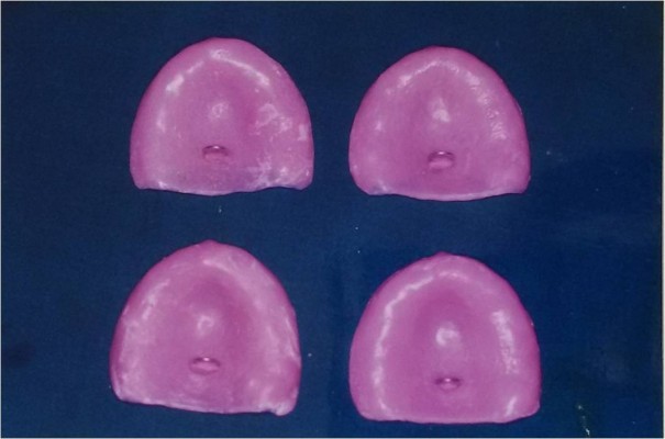
Trial closure was done, metal-to-metal contact was obtained. The packed flask was allowed to bench cure for 15 minutes. The polymerization was carried out under similar conditions for all flasks at a temperature of 74°C for 2 hours and at 100°C for 1 hour.7 After polymerization the flasks were allowed to bench cool for 1 hour. The denture bases were recovered carefully and finished. The four denture bases were checked for accuracy and that they were identical in all respects, except for their posterior palatal seal. The denture bases were marked “M”, “A”, “B”, and “C” respectively (Figure 5).
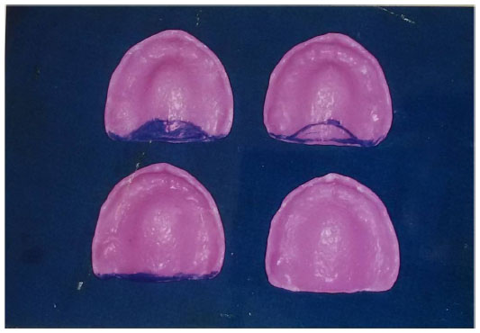 | Figure 5 Denture bases with different posterior palatal seal. |
The loop (hook) made of 19 gauge stainless steel wire was attached to the denture base with self-cured acrylic resin, exactly in the midline, the loops were placed 1 cm inwards to the anterior portion from the distal border of the denture base (Figure 6).
 | Figure 6 Denture bases with hook. |
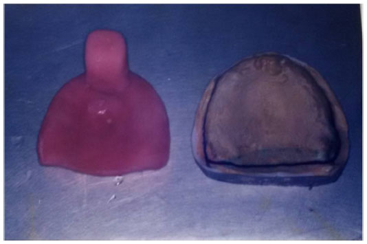
The denture bases “M”, “A”, “B”, and “C” were subjected to a retention test. Required force to dislodge bases was exerted and retention value was calculated for each denture base. After completing the test, the denture base “M” without posterior palatal seal was re-designated as “MO”, and used to develop the posterior palatal seal with functional method by tracing the seal with low fusing compound (Figure 7). Thus the five denture bases were marked as:
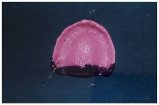 | Figure 7 Posterior palatal seal developed with low fusing compound. |
- M – without posterior palatal seal;
- A – single bead posterior palatal seal (scraped type);
- B – double bead posterior palatal seal (scraped type);
- C – butterfly shaped posterior palatal seal (scraped type);
- MO – posterior palatal seal with low fusing compound with functional method.
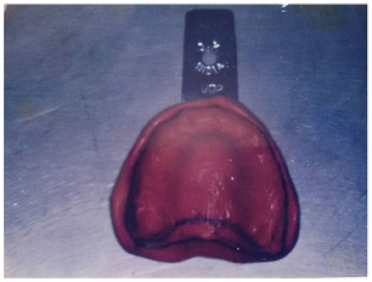
The five denture bases with different types of posterior palatal seals were subjected for evaluation of retention using a specialized T device in which a tipping force was developed extra-orally in a caudal direction to evaluate the retentive values of the denture bases (Figure 8).
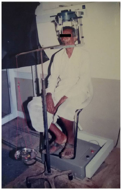 | Figure 8 Force applied on denture base using T-device. |
Results
In this study the force required to dislodge the maxillary denture bases was measured in grams. These forces were considered as the retentive values. For each of the ten patients, three readings were noted with each denture base. Thus, in all, fifteen readings were obtained from five denture bases. Therefore, in the study of ten patients a total of 150 readings were taken.
Analysis of variance (ANOVA) test was applied to compare the retention valves and Newman-Keuls student test for the simultaneous comparison.
A mean value was calculated for statistical analysis from each set of the three readings for each of the five denture bases. The mean retentive values are shown in Table 1 and graphically represented in Figure 9.
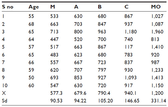 | Table 1 Mean retentive values in grams |
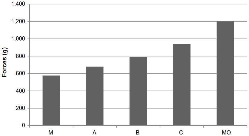 | Figure 9 Mean retentive values for different groups |
Retention of denture bases with different posterior palatal seals were compared with denture base “M” (without posterior palatal seal) and are shown in Tables 2 and 3. The retention values of significance between “A” and “C”, “MO”, “C” and “MO” (P<0.5); and those highly significant between “M” and “C”, “MO”, “B” and “MO” (P<0.01) can be seen.
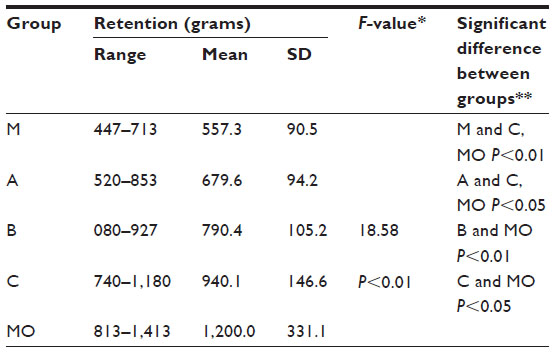 | Table 2 Comparison of retention between different types of posterior palatal seal |
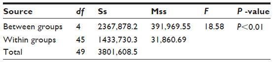 | Table 3 Analysis of variance |
The highly significant values are recorded with the denture base with butterfly shaped posterior palatal seal and functionally developed posterior palatal seal with low fusing compound.
Comparisons of retention between three different posterior palatal seals obtained with arbitrary scraping method are shown in Tables 4 and 5. It shows a significant difference between A–C (P<0.01) and B–C (P<0.05); and the differences between A–B were found to be insignificant.
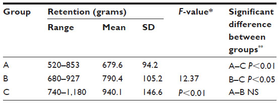 | Table 4 Comparison of retention between different posterior palatal seals obtained with arbitrary method |
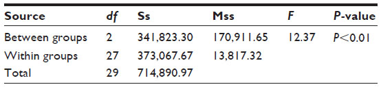 | Table 5 Analysis of variance for arbitrary method |
When comparing the scraped type of posterior palatal seals with each other, the butterfly shaped posterior palatal seal and double beaded posterior palatal seal showed highly significant retentive values.
Percentage increase in retention values of denture bases with different types of posterior palatal seals when compared to “M” (without posterior palatal seal) is shown in Table 6.
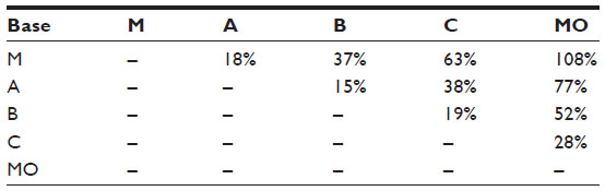 | Table 6 Percentage increase in retention from one group to another |
The results showed that there is a consistent increase in retention of denture bases with different types of posterior palatal seals when compared to denture bases without posterior palatal seals.
The posterior palatal seal obtained by functional method provided greater retention value than those obtained with the scraping method when compared with each other and with a denture base without a posterior palatal seal.
Discussion
The fact that posterior palatal seal plays an important role in enhancing the retention and stability of complete maxillary denture is well recognized.2,3 By providing intimate contact with the posterior palatal tissue, it reduces the gagging reflex. Further, it partly compensates for the palatal discrepancy occurring as a result of curing shrinkage of acrylic resins.4
The available literature shows that the displacement of the soft tissue is a critical factor in developing the posterior palatal seal.5 Skinner and Chung3 demonstrated the importance of posterior palatal seal experimentally. Several methods are used to develop the posterior palatal seal,1,6–13 namely in the arbitrary method, by scraping the cast at the distal border of the denture depending on the compressibility of the tissue. Hamrick12 stated that an upward extra-oral force to test the retention of maxillary denture was closer to a functional situation. Avant15 stated that in extra-oral caudally directed forces, some patients feel pain and discomfort in the anterior ridge. So he developed a “T” shaped device which could be used to apply tipping forces, where the anterior ridge acts as fulcrum. Antolino16 showed the application of forces to dislodge the denture at the posterior border of denture was closer to the dislodging pattern of denture during function.
The most obvious finding in the present study was that irrespective of the type of posterior palatal seal employed there occurred a consistent and substantial increase in the retention of denture base. Since the denture bases are similar to each other except for the posterior palatal seal. Retention was checked at constant leverage action, the salivary flow and the degree of mouth opening was maintained constant throughout the study. The variability in the retention value of the test bases could only be attributed to the varied situation of the posterior palatal seal.14
Comparison of retention between different types of posterior palatal seals shows significant to highly significant retentive values when compared to a denture base without a posterior palatal seal.
The findings of the present study were found to be at par with past studies made by other investigators. Mean retentive value of “M” was 577.3 grams against values of “A”, “B”, “C”, and “MO” bases, which were 679.6, 790.4, 940.1, and 1,200 grams respectively.
The percentage increase in retention of denture base when compared to denture base “M” showed an increased retentive value of 108%, 63%, 37%, and 18% with the denture bases “MO”, “C”, “B”, and “A”, respectively.
Conclusion
Within the limit of this in vivo study, it was concluded that the incorporation of posterior palatal seal is essential for obtaining optimum retention of maxillary complete denture. All types of posterior palatal seals evaluated provided a consistent and substantial increase in the retention of denture base. The retention of denture base varied with the type of posterior palatal seal.
Among the “scraped” type of posterior palatal seal evaluated, the “butterfly” shaped posterior palatal seal showed superior retention compared to “single bead” and “double bead” type of posterior palatal seal. The functional type of posterior palatal seal developed by the addition of low fusing compound provided higher retentive values when compared to the “scraped type” of posterior palatal seal.
Disclosure
The authors report no conflict of interest. It is self-funded research.
References
Hardy IR, Kapur KK. Posterior border seal – its rationale and importance. J Prosthet Dent. 1958;8:386–397. | |
Roydhouse RH. The retention of dentures. J Am Dent Assoc. 1960;60: 159–163. | |
Skinner EW, Chung P. The effect of surface contact in the retention of a denture. J Prosthet Dent. 1951;1(3):229–235. | |
Sykora O, Sutow EJ. Posterior palatal seal adaptation: influence of processing technique, palate shape and immersion. J Oral Rehabil. 1993;20(1):19–31. | |
Sidney SI. Dimensions and displacement patterns of the posterior palatal seal. J Prosthet Dent. 1971;25(5):470–488. | |
Dhir RC, Joneja OP. An evaluation of different types of posterior palatal seals in complete denture base retention. J Indian Dent Assoc. 1982;54(2):59–65. | |
Fish EW. Principles of Full Denture Prosthesis. 6th ed. Michigan: Staples Press; 1964. | |
Gesser HD, Castaldi CR. The preparation and evaluation of wetting dentures for adhesion and retention. J Prosthet Dent. 1971;25(3):236–243. | |
Giglio JJ, Lace WL, Arden H. Factors affecting retention and stability of complete dentures. J Prosthet Dent. 1962;12:848–856. | |
No authors listed. The Glossary of Prosthodontic Terms. J Prosthet Dent. 2005;94(1):10–92. | |
Green AJ, Harman L. Influence of diuretics on complete denture retention: a preliminary report. J Prosthet Dent. 1980;43(5):506–507. | |
Hamrick JE. A comparison of the retention of various denture-base materials. J Prosthet Dent. 1962;12:666–677. | |
Campbell RL. Some clinical observations regarding the role of the fluid film in the retention of dentures. J Am Dent Assoc. 1954;48(1):58–63. | |
Abdullah MA. Surface tension in retention of complete dentures. J Prosthet Dent. 1972;28(2):141–144. | |
Avant WE. A study of some factors associated with denture retention. J Prosthet Dent. 1973;29(4):383–389. | |
Antolion C, Kotwal K, Mangelsdorff D. Analysis of posterior palatal seal and the palatal form as related to the retention of complete dentures. J Prosthet Dent. 1982;47(10):23–27. | |
Avant WE. A comparison of the retention of complete denture bases having different types of posterior palatal seal. J Prosthet Dent. 1973;29(5):484–493. |
 © 2014 The Author(s). This work is published and licensed by Dove Medical Press Limited. The full terms of this license are available at https://www.dovepress.com/terms.php and incorporate the Creative Commons Attribution - Non Commercial (unported, v3.0) License.
By accessing the work you hereby accept the Terms. Non-commercial uses of the work are permitted without any further permission from Dove Medical Press Limited, provided the work is properly attributed. For permission for commercial use of this work, please see paragraphs 4.2 and 5 of our Terms.
© 2014 The Author(s). This work is published and licensed by Dove Medical Press Limited. The full terms of this license are available at https://www.dovepress.com/terms.php and incorporate the Creative Commons Attribution - Non Commercial (unported, v3.0) License.
By accessing the work you hereby accept the Terms. Non-commercial uses of the work are permitted without any further permission from Dove Medical Press Limited, provided the work is properly attributed. For permission for commercial use of this work, please see paragraphs 4.2 and 5 of our Terms.